

Some topics grab attention instantly, and digital anatomy is one of them. It’s not a buzzword. It’s not a passing trend. It’s the next big shift in how we see, study, and teach the human body.
What once felt locked inside textbooks and cadaver labs is now opening up through interactive 3D models, motion graphics, and 3D animation services that bring anatomy to life with unbelievable clarity.
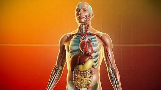
Medical students, educators, and healthcare companies are no longer limited to flat diagrams or static illustrations. They want movement, precision, and access to complex structures in a way that feels natural.
Digital anatomy delivers all of that with a mix of medical imaging and education technology, surgical simulation and training animation, and 3D anatomical structures that look and behave like the real thing.
At Prolific Studio, we’ve seen how fast this shift is happening. Schools and hospitals are turning to digital tools to fill gaps that traditional teaching can’t handle anymore. Brands in healthcare want 3D product video animation services to explain devices in a clean, visual way.
Even patients are benefiting because complex procedures are easier to understand through medical device animation and simple-to-follow visuals.
Digital anatomy is not just a tool. It’s a new lens for the entire healthcare industry.
[elementor-template id=”13845″]
What Digital Anatomy Really Means Today
Digital anatomy is the practice of studying and teaching the human body through interactive, digital, and often animated tools. This includes VR-based exploration, AR-assisted training, 3D-printed models, and high-fidelity simulations powered by professional medical animation services.
The idea sounds simple. The impact is not.
For decades, the cornerstone of medical education revolved around cadavers, physical models, and thick atlases. Those tools still matter, but they have limits. Cadaver access is low in many regions. Students struggle with translating 2D illustrations into spatial understanding. Physical models rarely capture variations in anatomy or show the dynamic behavior of organs.
Digital anatomy fills those gaps by giving access to:
- interactive 3D models
- medical animation services
- 3D anatomical structures
- surgical simulation and training animation
- 3D-printed models
- motion graphics that simplify complex systems
This shift is pulling medical students closer to the kind of learning they always wanted: clear, visual, and repeatable anytime.
The Growth of Digital Anatomy in Medical Learning
Students today expect consistency, clarity, and flexibility in how they learn. Digital anatomy gives them the freedom to replay movements, zoom into structures, peel back layers, and study functions that would be impossible to observe in a formal lab.
It’s reshaping medical education in several ways:
Interactive 3D models are becoming the new standard
These models respond in real time. Rotate them. Isolate a muscle. Explore a nerve pathway. Add motion graphics to see how a joint actually moves. This kind of clarity wasn’t available in the past.
3D medical education is more practical
Anatomy blends with physiology, pathology, and medical imaging. Students can match what they see in a 3D model with CT or MRI scans. It sharpens clinical reasoning early and reduces confusion that often appears during internships.
Training for medical scenarios is stronger
Surgical simulation and training animation allow learners to practice procedures repeatedly without risk. They can make mistakes, adjust, and try again.
Digital anatomy keeps learning active, not passive.
How Video Animation Agencies Are Pushing Digital Anatomy Forward

Behind many of these innovations are creative studios like Prolific Studio. A reliable video animation agency isn’t just an artist team anymore. It’s a technical partner that blends storytelling with science.
Medical educators, device companies, and hospitals now depend on visual material to communicate with clarity. They want work that is accurate but also appealing enough to hold interest. That’s pushing animation teams to deliver:
- lifelike motion graphics
- detailed medical animation services
- realistic textures for 3D anatomical structures
- training videos that match real surgical procedures
- 2D vs 3D animation comparisons for education
- voice generation in animation for narration and lectures
The demand isn’t slowing down. Each year, more institutions request deeper visualization tools. This trend is becoming one of the top animation trends in healthcare content, and it’s clear why: when anatomy looks real, learning improves instantly.
A Look at the Evolution of Digital Anatomy
Digital anatomy didn’t appear out of nowhere. It evolved through decades of medical imaging and education technology, advancing step by step.
Early medical sketches sparked it all
Physicians relied on handmade drawings. Helpful, yes, but very limited.
Cadaver labs filled a huge gap
They offered real texture, layers, and variation. Still essential today, but not always accessible.
Medical imaging changed everything
The advent of medical tools like X-rays, MRI, and CT scans revealed structures inside a living body. A major step forward in accuracy.
3D modeling pushed anatomy into a new form
Once computers could reconstruct slices from MRI or CT into 3D shapes, anatomy finally gained depth on a screen.
VR, AR, and MR opened an entirely new form of teaching
Students could walk through organs, study large structures in motion, and merge physical and digital learning.
This timeline reflects a natural shift. Each stage brought anatomy closer to how the human body actually behaves. Digital anatomy completes the picture.
Modern Tools Behind Digital Anatomy
Digital anatomy stands on a set of powerful technologies that make exploration both accurate and intuitive.
High-quality 3D modeling
Accurate 3D anatomical structures start with clean imaging data. Then, artists and medical experts build models that highlight layers, variations, and tissue behavior. These models are the core of 3D medical education.
Immersive VR environments
Wearing a VR headset places students inside a fully interactive version of the human body. They aren’t observing anatomy, they’re inside it.
Augmented reality for on-the-spot learning
AR brings anatomy into classrooms or clinics through digital overlays. It helps medical staff explain conditions directly on a patient’s body outline.
Mixed reality for flexible study
MR blends real and digital elements. It helps teams collaborate, study 3D models together, and plan procedures with better clarity.
3D-printed models for hands-on training
Even in a digital world, 3D-printed models offer the tactile experience students and surgeons often miss. They help in surgical planning, device testing, and patient education.
Motion graphics and medical animation
These tools break down complex body functions into simple, flowing visuals. Perfect for explaining processes that are too fast, too small, or too hidden to see in real life.
Digital anatomy thrives on this mix of tools because each solves a different problem in medical training.
Why Medical Companies Prefer 3D Product Video Animation Services
Healthcare brands need clarity. Their devices and solutions are often technical and hard to explain verbally. That’s why 3D product video animation services have become a major part of their marketing and training material.
These animations make it easier to show:
- implant mechanisms
- device pathways
- step-by-step procedures
- internal movements
- device interactions with human tissue
Clear visuals help doctors trust a product faster. They also help buyers, investors, and patients understand exactly how something works.
This is one reason medical animation services are growing fast and becoming a core part of digital anatomy education, too.
Why Digital Anatomy Matters for Surgeons and Clinical Teams

Digital anatomy isn’t only a tool for students. Surgeons use it daily for planning, testing ideas, and explaining procedures to patients. A simple conversation with a patient often turns into a clearer experience when paired with a short visual created through medical animation services.
Instead of long explanations, they show a quick animation of the exact joint, nerve, or organ. The message lands instantly.
Some surgeons use interactive 3D models to practice the trickiest parts of a procedure. These models reveal curves, thickness, and depth with precision. When combined with surgical simulation and training animation, they get a clear picture of how tools behave inside the body. It reduces stress and builds confidence before entering the operation room.
Digital anatomy has become part of pre-op planning, team discussions, and patient counseling. It removes the guesswork and replaces it with accuracy that comes straight from medical imaging and education technology.
Why Digital Anatomy Beats Traditional Methods in Key Areas
Old methods still have value, but digital anatomy provides consistency that physical resources can’t match. Students across different campuses learn from the same accurate source. No distractions. No damaged cadavers. No faded illustrations.
Accuracy stays intact
A model created once through 3D animation services remains clean forever. No wear and tear. No missing parts.
Repeatability for every learner
Digital tools allow unlimited reviews. Pause it. Rewind it. Go frame by frame. Each student learns at their own pace.
Scalability for huge classes
A class of 250 students can explore the same model at the same time. Nobody gets left behind.
These advantages bring digital anatomy to the forefront of reshaping medical education.
Why 2D vs 3D Animation Still Matters in Healthcare Education
The debate between 2D vs 3D animation continues, especially in medical education. Both have strengths, but digital anatomy thrives mostly on 3D because anatomy requires depth.
2D animation fits simple explanations
For surface-level processes like blood flow diagrams or patient education, 2D offers clarity and speed.
3D animation fits detailed anatomy
When a topic demands spatial understanding, joint rotation, organ movement, and device placement, 3D becomes essential.
Hybrid animation blends both worlds
Some lessons work better when 2D motion graphics are combined with 3D models. This mix creates smooth storytelling without overwhelming the viewer.
Digital anatomy bends easily in all directions, giving instructors and animation teams total control.
How Digital Anatomy Helps Medical Students Learn Faster

Medical textbooks carry detail, but they can’t show movement. Students often memorize diagrams only to forget how structures behave in real life. Digital anatomy fixes that.
Clear visual memory
When students watch a 3D animation of a beating heart or a muscle contraction, the detail sticks. The brain holds onto visuals more easily than long descriptions.
Stronger recall during exams
Students who train with 3D anatomical structures are more comfortable identifying variations. Their answers come faster because they’ve seen structures from angles that books never offer.
Better understanding of complex regions
Areas like the inner ear, cranial nerves, and pelvic floor are notoriously confusing. Motion graphics, VR-based layers, and 3D-printed models solve that frustration.
Digital anatomy removes the fog and brings students straight into the experience.
Frequently Asked Questions
How do interactive 3D models help medical students?
They let students view structures from every angle, peel layers, and understand movement that books cannot show.
Are digital anatomy tools accurate?
Accuracy comes from medical imaging and education technology such as MRI, CT, and ultrasound scans, combined with expert modeling from medical animation services.
Do surgeons rely on digital anatomy?
Yes. Surgeons use surgical simulation and training animation for planning and revisiting complex procedures.
How does digital anatomy support device companies?
3D product video animation services show precise device function, making sales, training, and patient education faster and easier.
Is 2D animation still used in medical content?
Yes. It works well for simple educational videos. For complex anatomy, 3D is preferred.
Can digital anatomy help patients understand procedures?
Clear medical device animation helps patients visualize what will happen during surgery or treatment. It builds trust and reduces anxiety.
Final Words
Digital anatomy isn’t optional anymore. It’s becoming the backbone of medical learning, device training, and clinical communication. Students expect visuals that match the complexity of the body. Surgeons want precise planning tools. Healthcare companies need animation that explains products without confusion. Clinics need patient-friendly visuals.
Prolific Studio stands at the front of this shift with professional medical animation services, interactive 3D models, and complete 3D animation services designed for modern healthcare teams. If your institution wants stronger training, better patient education, or clear device explanations, digital anatomy is the fastest way to get there.
When you’re ready to bring clarity, accuracy, and visual power into your medical content, Prolific Studio can build it.
Related Articles:


